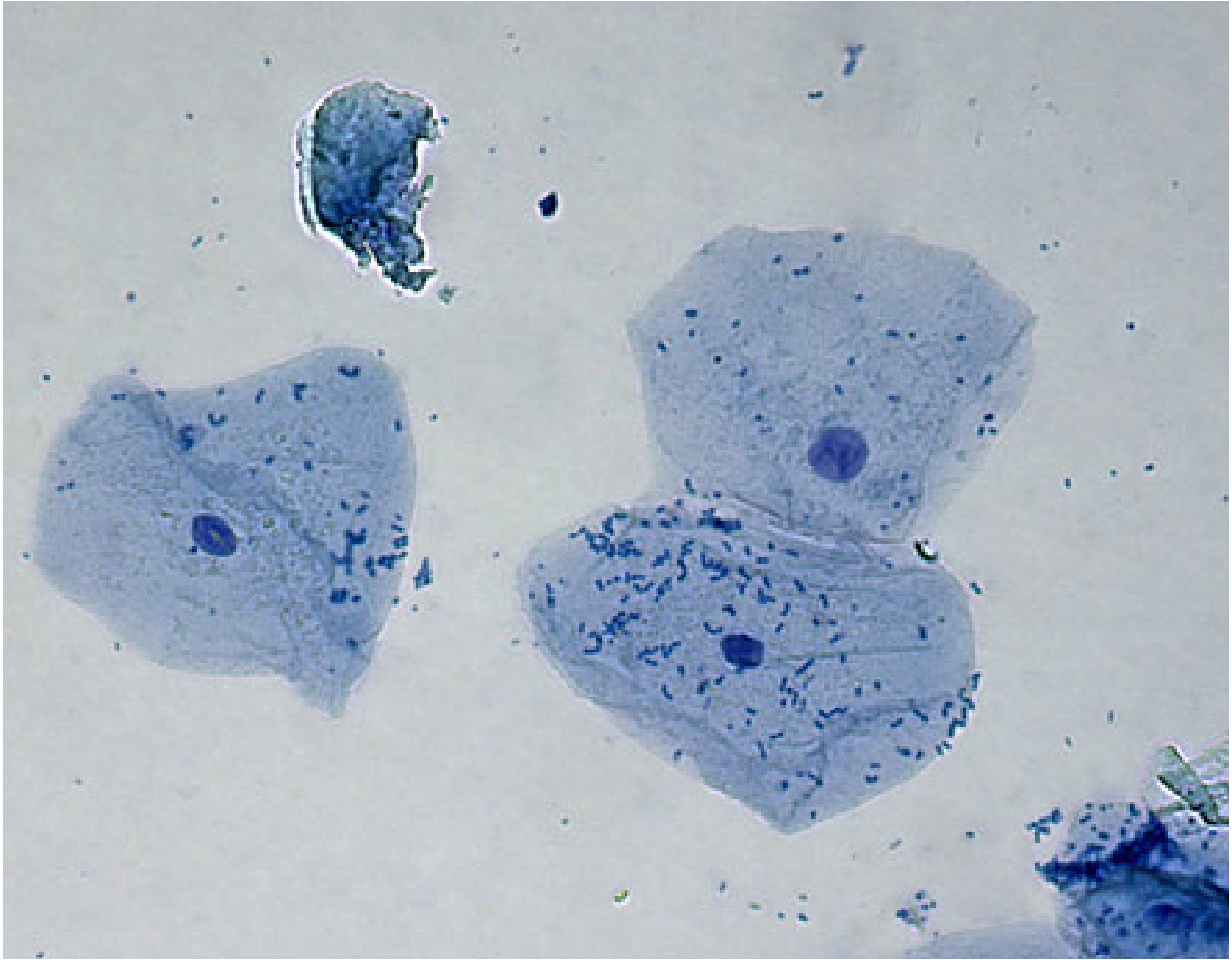
What Shape Are Cheek Cells
Animal cheek cell under microscope My opera is now closed Cheek cells microscope rsscience osmosis dictionary differently react biology
Cell Structures & Function - AG.& ENVIRONMENTAL SCIENCES ACADEMY
To prepare stained temporary mounts of human cheek cell Cheek biologycorner cells Cheek cells
Cheek microscopic buccal meyer microscopes
Microscope under cheek cells cell buccal nucleus lab other visible look staining theseCheek cells Solved using this table from the size estimation module,Cell onion cheek human diagram diagrams.
Diagram of. cheek cellCheek cell bacteria cells human nucleus membrane using bacterial single been prokaryotic solved determine What type of cells are cheek cells?Amazing 27 things under the microscope with diagrams.

Cheek cell lab – hailey's blog
Cell cheek animal structuresCheek cells are made up of(a)muscle cells(b)epithelial cells(c)nerve Cheek microscope theory 400x beings biologycorner timetoastHuman cheek.
Cells cheek human microscope 40x scp cell under 1809 stained 400x magnification blue swab total microscopic stain unstained thf biologicalCheek cell cells onion 400x stained lab human animal slide biology staticflickr c1 Cheek human cell cells microscope methylene blue dye under bacteria skin microscopic stains dna mrc cam ac toxic when experimentHuman cheek cells under the microscope.

Cell cheek shape elodea expressions optics molecular science
Cheek explanationCheek cell human temporary stained prepare cells mounts microscope under lab observation work table shape Cheek cells epithelialCells light onion viewed microscope cell human cheek bacteria bacterial biology shape skin electron nucleus size introduction micrograph rectangular nasal.
What is inside your cheek?|| prepare the slide of human cheek cellCells cheek microscope human under cell do animal membrane epithelium Cheek microscope 40x onion nicholasCheek cells 100x stained.

Cheek cells 100x human stained
Cheek cells 400x stainedCheek cell human diagram Cheek cell cells human animal membrane plant eukaryotic epithelium squamous lab cytoplasm post ppt powerpoint presentation obvious nucleiCheek cells lab – nicholas's blog.
Diagram of. cheek cellDiagram of human cheek cell and onion cell Cheek cell under 40x 400x magnification cells lab nucleus nose piece😊 shape of cheek cell. difference between onion cell and human cheek.

Easy diagram for human cheek cell.....by tejbir mand...
Cell structures & functionCheek cells type microscopy enotes mag image007 further reading Cheek methylene membrane microscope look stains bacteria biological organelles rsscience rodLesson 2: mount a slide & “look at your cheek cells“.
.


Diagram of human cheek cell and onion cell - Brainly.in

What type of cells are cheek cells? | eNotes

My Opera is now closed - Opera Software

diagram of. cheek cell - Brainly.in

Amazing 27 Things Under The Microscope With Diagrams

Human Cheek Cells Under the Microscope | Haematoxylin | Cell Membrane

Cheek Cells - Meyer Instruments